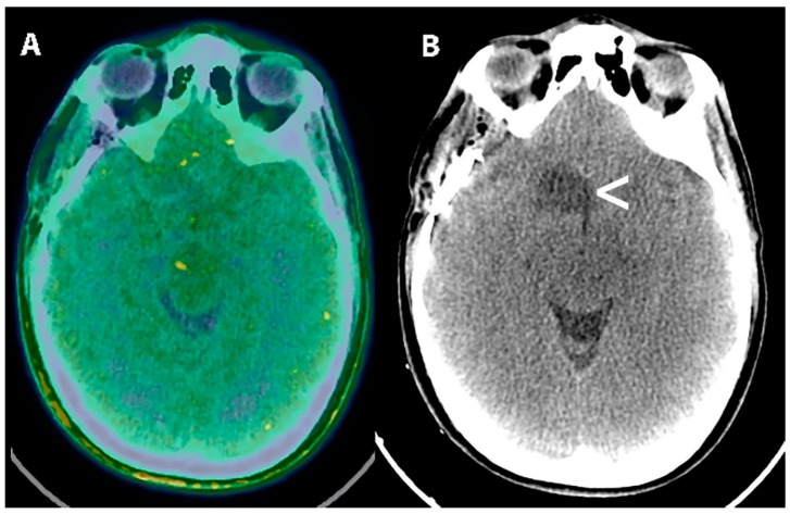
Figure 5.
[18F] FDOPA PET (A) and T2 FLAIR MRI (B) in a 14-year-old female patient with the suspect of primary brain tumor. T2 FLAIR image showed an area of mild hyperintensity in the left temporal lobe(B,<). [18F] FDOPA PET/CT scan demonstrated increased uptake of the radiopharmaceutical in the lesion with a Standardized uptake value equal to 2.2 vs. 1.1 of the surrounding brain tissue (A,<). The patient was surgically treated and the subsequent diagnosis of low-grade glioma (WHO grade II).

