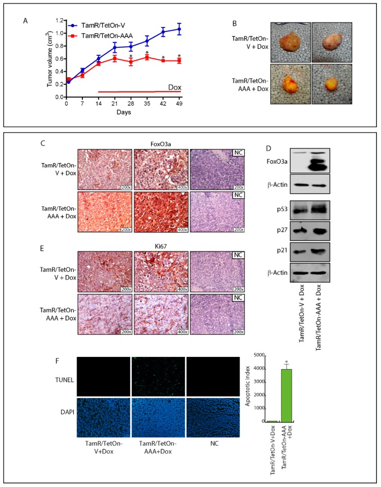
Figure 4.
FoxO3a over-expression inhibits the growth of engineered TamR/TetOn derived tumor xenografts. (A) TamR/TetOn-V and TamR/TetOn-AAA cells were injected subcutaneously in female nude mice (see Materials and Methods). When the tumors reached average ~0.2 cm3, mice were treated with Doxycycline hyclate (Dox) for 35 days. Tumor growth was monitored by caliper, measuring the visible tumor sizes at indicated time points. Data represent the mean ± SD of measurements. * p < 0.05 vs. TamR/TetOn-Vector xenografts. (B) At the end of the experiment, tumors were explanted and representative tumor images are shown. The expressions of FoxO3a (C) and Ki-67 (E), as well as the apoptotic index (TUNEL assay) (F) were evaluated in tumor sections deriving from Dox-treated mice injected with engineered TamR/TetOn cells. NC: negative control. (D) An amount of 50 µg of lysates from explanted tumors were subjected to WB analysis to evaluate Dox-induced FoxO3a overexpression. β-actin was used as the loading control.

