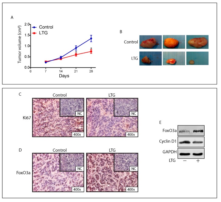
Figure 6.
LTG induces FoxO3a and inhibits the growth of TamR derived tumor xenografts. (A) Xenografts were established with TamR cells in female mice implanted with E2 and successively, tamoxifen pellets (see Materials and Methods for details). One group was treated with 20 mg/kg/day LTG (n = 5) and a second group with vehicle alone (n = 5). Tumor mass was measured at indicated time points with a caliper. Data represent the mean ± SD of measurements. * p < 0.05 vs. control xenografts. (B) Representative images of explanted tumors at day 28. Tumor sections from mice at 28 days were formalin fixed, paraffin embedded, sectioned, and immunostained with hematoxylin and eosin Y (H&E) or incubated with antibodies directed against the epithelial marker cytokeratin 18 (Cyt 18) (Figure S9). Immunostaining was also performed for the proliferation marker Ki-67 (C) and for FoxO3a (D). (E) FoxO3a and CyclinD1 expression was assessed in protein extracts from xenografts excised from control mice and LTG treated mice. GAPDH was used as the loading control.

