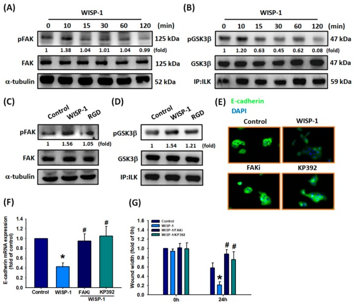
Figure 4.
Involvement of FAK and ILK in WISP-1-regulated EMT functioning. (A,B) SCC4 cells were incubated with WISP-1 (30 ng/mL) for the indicated time intervals; FAK and ILK activation was examined by Western blot assay. (C,D) Cells were pretreated with RGD (100 nM) for 30 min then treated with WISP-1 for 10 min; FAK and ILK activation was examined by Western blot assay. FAK and GSK3β content was used to normalize for pFAK and pGSK3βlevels. (E,F) Cells were pretreated for 30 min with a FAKi (10 M) and KP392 (10 M), prior to incubation with WISP-1 for 24 h. E-cadherin expression was examined by IF and qPCR assays. (G) Cells were pretreated for 30 min with a FAKi (10 M) and KP392 (10 M), then incubated with WISP-1 for 24 h. Cell migration was examined by the wound healing assay. Results are expressed as the mean ± SEM. * p < 0.05 compared with controls; # p < 0.05 compared with the WISP-1-treated group.

