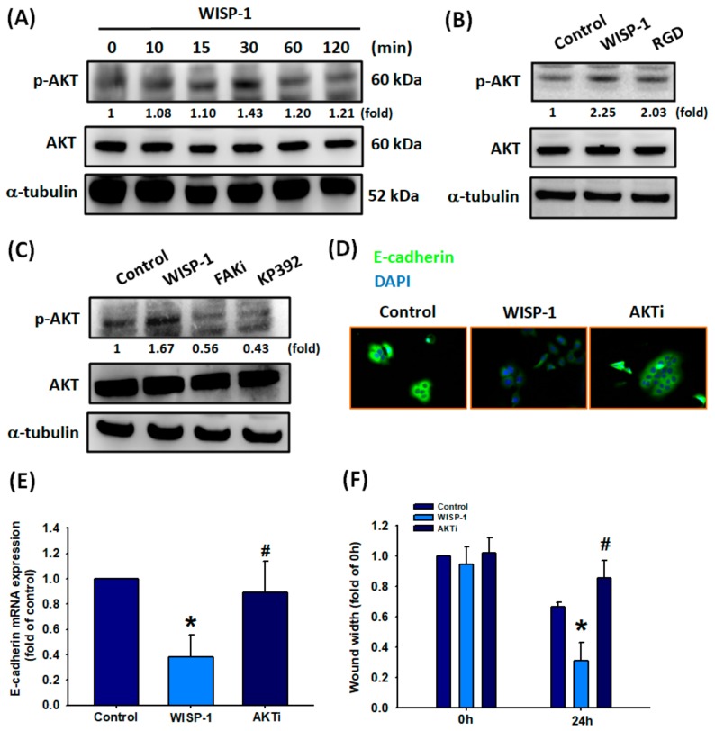
Figure 5.
WISP-1 induces Akt phosphorylation via the integrin αvβ3/FAK/ILK signaling pathway. (A) SCC4 cells were incubated with WISP-1 (30 ng/mL) for the indicated time intervals; levels of Akt phosphorylation were examined by Western blot assay. (B,C) Cells were pretreated with RGD (100 nM), a FAKi (10 M), or KP392 (10 M) for 30 min, then incubated with WISP-1 for 30 min. Akt activation was examined by Western blot assay. Akt protein was used to normalize for pAkt levels. (D–F) Cells were pretreated for 30 min with Akti (10 M), prior to incubation with WISP-1 for 24 h. E-cadherin expression was examined by IF and qPCR assays. Cell migration was examined by the wound healing assay. Results are expressed as the mean ± SEM. * p < 0.05 compared with the control group; # p < 0.05 compared with the WISP-1-treated group.

