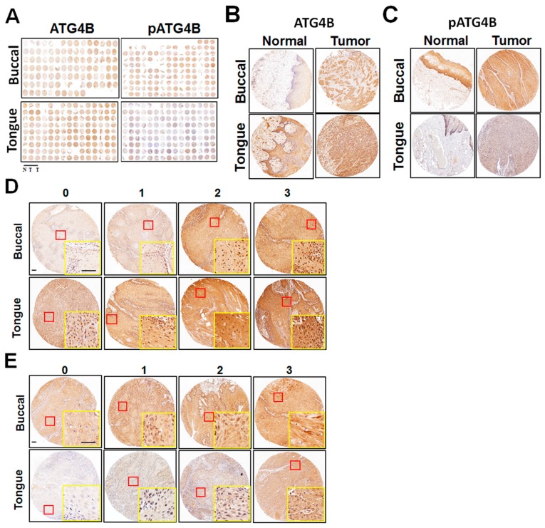
Figure 1.
Protein levels of ATG4B and phospho-Ser383/392-ATG4B in oral squamous cell carcinoma (OSCC). (A) Tissue microarrays consisting of tissues from 127 buccal mucosal SCC (BMSCC) patients and from 201 tongue SCC (TSCC) patients. Each sample for each patient included one portion of adjacent normal tissue (N) and two portions of tumor tissues (T). Tissue microarrays were stained via immunohistochemistry using antibodies against ATG4B or phospho-Ser383/392-ATG4B (pATG4B). Representative images are shown. (B) Representative immunohistochemistry staining of ATG4B or (C) phospho-Ser383/392-ATG4B (pATG4B) for paired tumor and adjacent normal tissues from BMSCC and TSCC. (D) The staining intensity for ATG4B or (E) phospho-Ser383/392-ATG4B (pATG4B) was categorized into four different levels, as the standard slides show: 0 = negative staining; 1 = weak; 2 = moderate; 3 = strong. Yellow rectangle is zoom in from red rectangle. Scale bar for (D) and (E): 100 μm.

