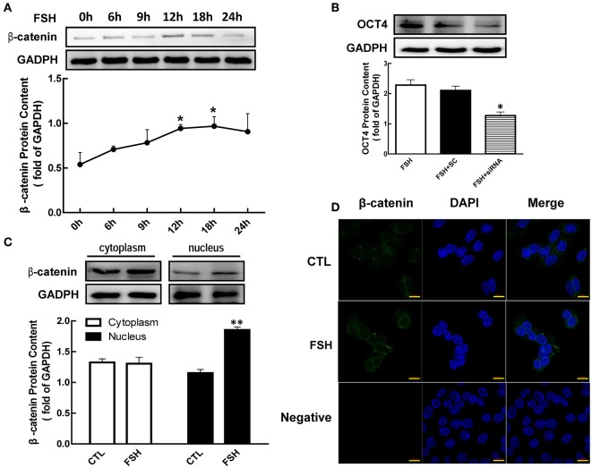
Figure 3.
Role of β-catenin in FSH-induced OCT4 expression. (A) Granulosa cells were cultured with FSH for different duration, and β-catenin expression was assessed by western blotting analysis. (B) Granulosa cells were transfected with β-catenin siRNA (scrambled sequence as control, SC) for 48 h using Lipofectamine 3000, and then treated with FSH for another 24 h. OCT4 protein was assessed by western blotting analysis. (C) Granulosa cells were cultured with FSH for 18 h, and β-catenin expression in the cytoplasm and nucleus were assessed. (D) FSH induced the shift of β-catenin distribution to the nucleus, which was detected by immunofluorescence. Data are presented as mean ± SEM of three independent experiments. *P < 0.05; **P < 0.01 compared with 0 h (A), FSH alone (B), or control (CTL, C), respectively. Bar, 10 μm.

