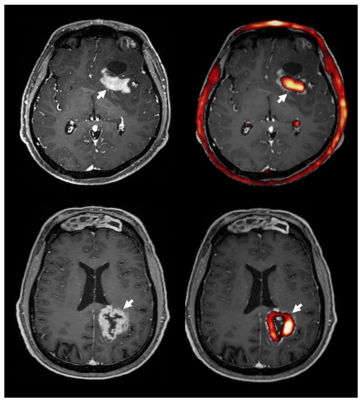
Figure 5.
The 18F-FMC uptake (right, red) and contrast enhancement on MRI (left) in two patients with GBM. Overlaying 18F-FMC PET onto post-contrast T1 weighted MRI showed that areas of contrast enhancement had a high 18F-FMC-uptake (white arrows). Overall, an average 94% of contrast enhancing regions had a 18F-FMC-uptake above the threshold, which was defined as three times that in the contralateral white matter. Note the normal high uptake in the choroid plexus and bone marrow.

