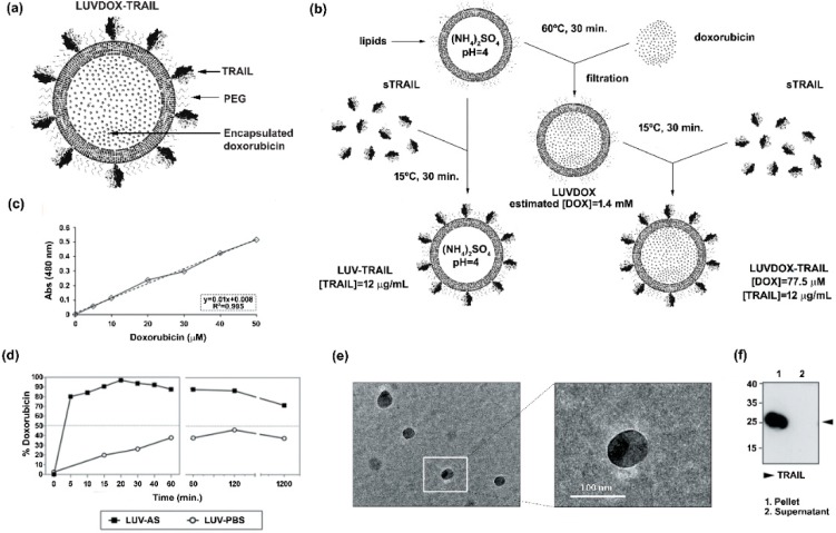
Figure 1.
(a) Schematic representation of large unilamellar vesicle doxorubicin-TNF-related apoptosis-inducing ligand (LUVDOX-TRAIL (LDT)). (b) Schematic illustration of the generation of LDT. Generation of LDT was performed as describe in the Experimental Section. (c) Standard curve of doxorubicin concentration. Absorbance at 480 nm of the indicated concentrations of doxorubicin (DOX) was measured. The DOX standard curve was used to interpolate the results obtained in (d). (d) Encapsulation efficiency and release profile of doxorubicin in LUV-AS. DOX was incubated with LUV-AS at a molar ratio of 1:3.8 (drug:lipid) for 30 min at 60 °C. At the indicated times, samples were taken and free DOX was separated from the liposomal fraction. Encapsulated DOX was quantified by measurement of absorbance at 480 nm. Efficiency is represented as percentage of total doxorubicin present in the liposomal fraction in every time point. (e) Scanning electron microscopy of LDT was carried out as described in the Experimental Section. Original magnification was 45,000×. (f) Coupling efficiency of soluble TRAIL in the LDT formulation. Once LUVDOX-TRAIL were generated, they were ultra-centrifuged at 60,000 rpm for 5 h, collecting the supernatant and resuspending the pellet. Aliquots from the pellet and the supernatant fractions were separated by SDS-PAGE and the presence of TRAIL in both fractions was assessed by Western blot.

