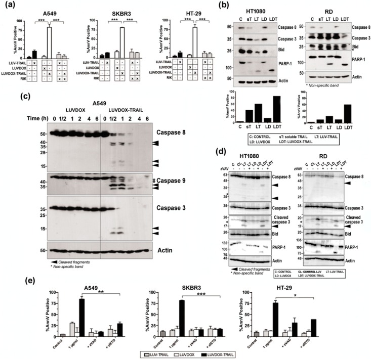
Figure 3.
(a) Roles of DOX and TRAIL in LUVDOX-TRAIL (LDT) cytotoxicity. A549, SKBR3, and HT-29 cells were treated with the indicated combinations of LUV-TRAIL (LT), LUVDOX (LD), and LDT (TRAIL at 1000 ng/mL). When indicated, cells were pre-treated with the TRAIL-blocking antibody RIK. Cell death was assessed by annexin-V staining after 6 h of treatment. Graphs show the mean ± SD of at least four experiments. *** p < 0.001. (b) Analysis of caspase activation after treatment with different TRAIL versions. HT-1080 and RD cells were untreated (Control, designed as C), or treated with soluble TRAIL (ST), LT, LD, and LDT at 1000 ng/mL. After 24 h, cells were lysed, and lysates were subjected to SDS-PAGE and to Western blot analysis. Levels of caspase-8, caspase-3, Bid, and PARP-1 were analyzed using specific antibodies. The level of actin levels was used as a control for equal protein loading. Cell death was quantified in parallel by flow cytometry after annexin-V staining (bottom panels). (c) Analysis of time-course caspase activation with LD or LDT. A549 cells were treated with LD or LDT (1 μg/mL TRAIL; 64.56 μM DOX) at the indicated times. Finally, cells were lysed, and lysates were subjected to SDS-PAGE and to Western blot analysis. (d) Analysis of caspase activation after treatment with different TRAIL versions. HT-1080 and RD cells were untreated (Control, designed as C), or treated with LUV alone (CL), LT, LD, and LDT at 1000 ng/mL. When indicated, cells were pre-treated with the pan-caspase inhibitor z-VAD-fmk (30 μM). After 24 h, cells were lysed, and lysates were subjected to SDS-PAGE and to Western blot analysis. Levels of caspase-8, caspase-3, Bid, and PARP-1 were analyzed using specific antibodies. Actin levels was used as a control for equal protein loading. (e) Role of caspases in LDT cytotoxicity. A549, SKBR3, and HT-29 cells were treated with LT, LD, or LDT (TRAIL at 1000 ng/mL). When indicated, cells were pre-treated either with the pan-caspase inhibitor z-VAD-fmk (30 μM) or with the specific caspase-8 inhibitor z-IETD-fmk (30 μM) for 1 hour. Cell death was assessed by annexin-V staining after 6 h of treatment. Graphs show the mean ± SD of at least four experiments. * p < 0.05, ** p < 0.01, *** p < 0.001.

