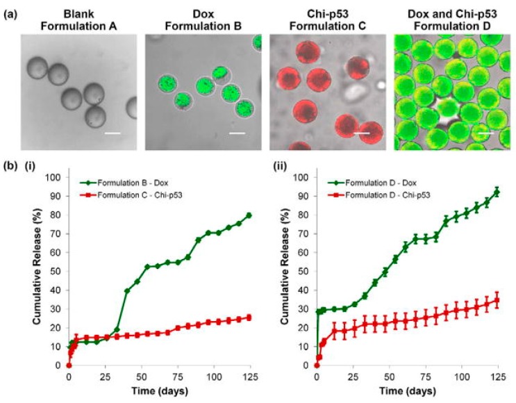
Figure 6.
(a) Transmitted light and laser scanning confocal (overlay) micrographs of blank and drug loaded double-walled PLLA (PLGA) microspheres. The distribution of DOX in Formulations B and D microspheres is indicated in green. The distribution of chi-p53 NPs in formulations C and D microspheres is indicated in red and yellow (colocalization of red and green), respectively. Scale bar = 50 μm. (b) In vitro DOX and chi-p53 release from double-walled PLLA(PLGA) microspheres. Reprinted with permission from Elsevier, Xu et al., Biomaterials, 2013 [58].

