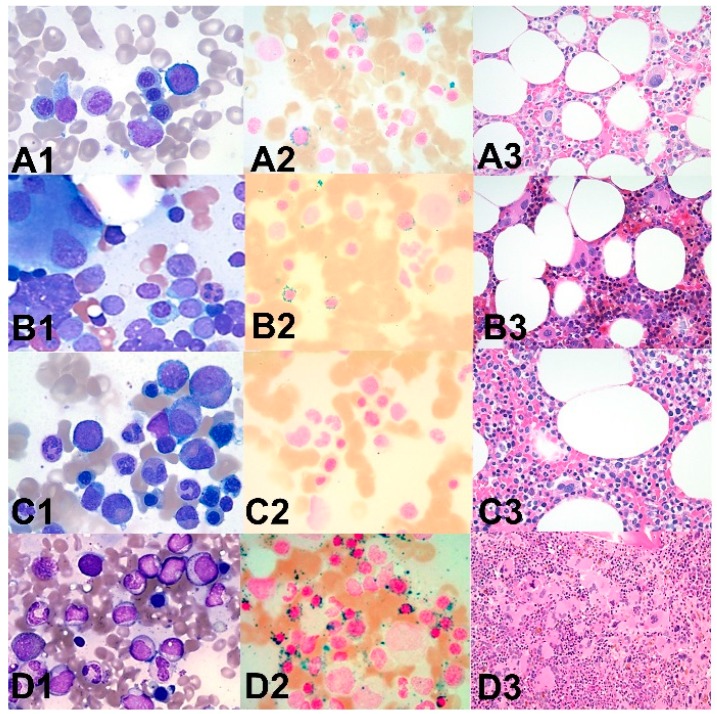
Figure 3.
Morphologic features of representative patients with mutations in splicing factors. Bone marrow aspiration smears (1: Wright–Giemsa stain, ×500) and iron stains (2, ×400) and core biopsies (3: H&E stain, ×200) in representative cases with myeloid malignancies and mutation in splicing factor genes. A1–3: A patient with myelodysplastic syndrome with ring sideroblasts and multilineage dysplasia with SF3B1 and ZRSR2 mutation (detected by next generation sequencing) showing mild dyserythropoiesis (A1), increased ring sideroblasts (A2) and dysmegakaryopoiesis (A3). B1–3: A patient with acute myeloid leukemia with SF3B1 mutation showing increased myeloblasts and dysmegakaryopoiesis (B1,B3) and increased ring sideroblasts (B2). C1–3: A patient with myelodysplastic syndrome with U2AF1 mutation showing minimal dyserythropoiesis and no increased blasts (C1), no increased ring sideroblasts (C2) and mild dysmegakaryopoiesis (C3). D1–3: A patient with myelodysplastic/myeloproliferative neoplasm with thrombocytosis and ring sideroblasts with SF3B1 mutation showing no significant dysplasia in granulocytes and erythroid cells (D1), increased ring sideroblasts (D2) and dysmegakaryopoiesis (D3).

