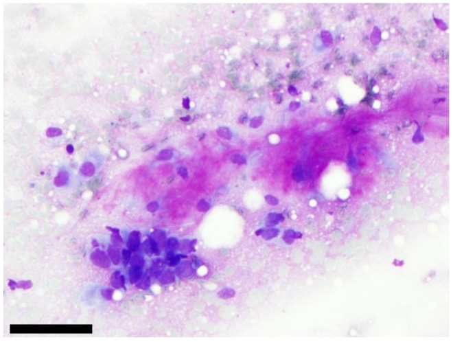
Figure 2.
Example of one case classified as SUMP with unspecified features after cytological analysis and diagnosed as poorly differentiated carcinoma after surgery. On this picture, we can see a cluster of atypical cells with high nuclear/cytoplasmic ratio which is close to a fibrillary matrix (MGG staining). FISH with PLAG1 or MYB probes were negative, (scale bar: 100 µm).

