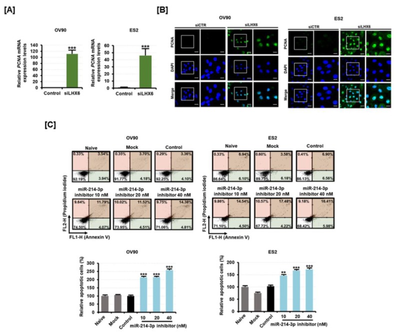
Figure 5.
Effect of miR-214-3p inhibitor and LHX6 knockdown on the apoptosis of OV90 and ES2 cells. (A) The expression of proliferating cell nuclear antigen (PCNA) mRNA was measured by qPCR analysis from the RNA of OV90 and ES2 cells transfected with control or siLHX6 (40 nM). (B) PCNA protein was detected (green) and the nucleus was counterstained with DAPI (blue) in OV90 and ES2 cells transfected with 40 nM of siLHX6 for 48 h. (C) Flow cytometric detection of apoptosis in OV90 and ES2 cells transfected with miR-214-3p inhibitor (20 nM, 40 nM, and 80 nM). Annexin V and propidium iodide (PI) fluorescence values were estimated by flow cytometry. The percentage of apoptotic cells (upper right and lower right quadrants) was analyzed in comparison to the control. All experiments were performed in triplicate. The asterisks indicate the significance compared to the control (*** p < 0.001 and ** p < 0.01). The scale bar represents 40 μm (in the first and third vertical panels) or 20 μm (in the second and fourth vertical panels).

