Figure 1.

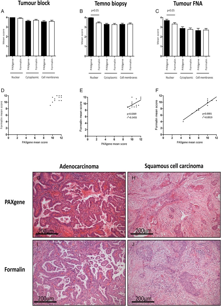
H&E staining and histomorphology. Histomorphology of PFPE and FFPE H&E stained sections of tumour block (A), Temno biopsy (B) and tumour FNA clot preparations (C). Nuclear staining of Temno biopsies and tumour FNA specimens was preferable in PFPE samples (both p < 0.05). Scores for nuclear, cytoplasmic and cell membranous staining were combined for tumour blocks (D), Temno biopsies (E) and for tumour FNA samples (F) and correlations between FFPE and PFPE scores were observed for Temno biopsies (r 2 = 0.3455, p = 0.0269) and tumour FNA samples (r 2 = 0.8919, p < 0.0001). Representative H&E staining of PFPE adenocarcinoma (G) and PFPE squamous cell carcinoma (H) and FFPE adenocarcinoma (I) and FFPE squamous cell carcinoma (J).
