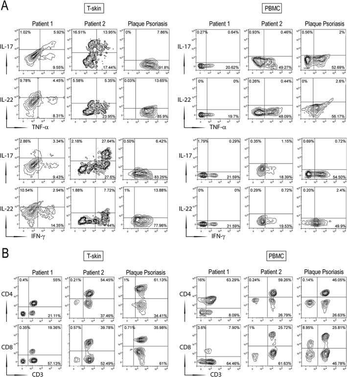
Figure 4.

Immunophenotypic characterization of skin T cells (T‐skin) and PBMCs isolated from HS patients with psoriasiform lesions. T‐skin cells (left panel) and PBMCs (right panel) were isolated from biopsies and blood, respectively, of patients 1 and 2 and from psoriatic patients (n = 2). Co‐expression of IL‐17, IL‐22, TNF‐α, or IFN‐γ on gated CD3+ cells (A), and surface CD4, CD8, and CD3 (B), were analyzed by flow cytometry. The percentage of positive fluorescent cells is shown in each quadrant. The results show the mean values of data obtained for one representative experiment out of three experiments.
