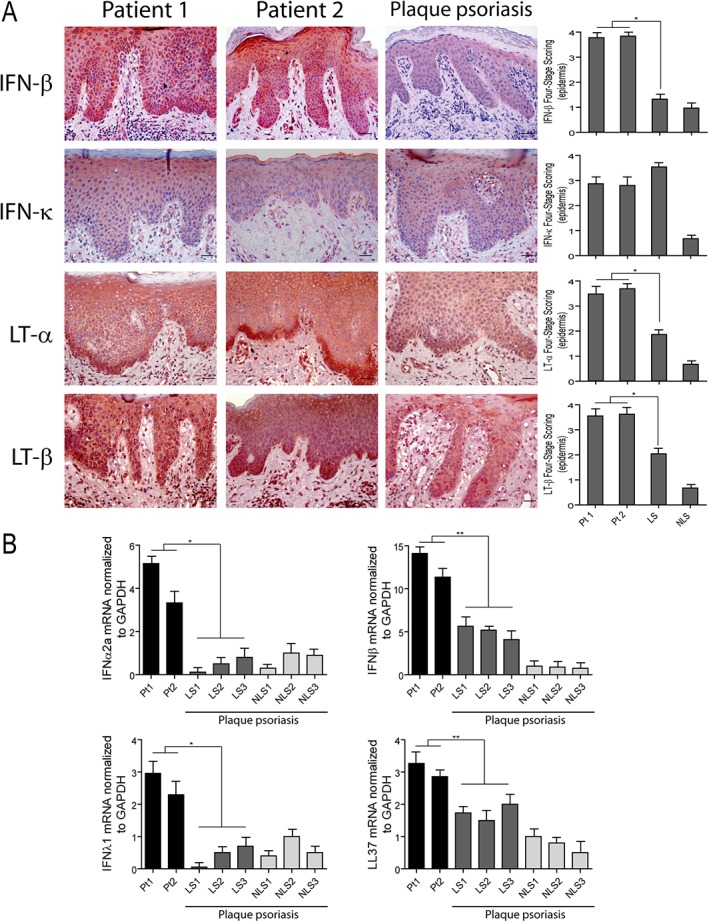
Figure 5.

Innate immunity molecules are overexpressed in the skin of HS patients after TNF‐α treatment. (A) Immunohistochemistry analysis of paradoxical skin reactions obtained from patients 1 (Pt1) and 2 (Pt2) shows an increase of IFN‐β, LT‐α, LT‐β, and similar IFN‐κ positivity, when compared with psoriatic skin lesions. LS and NLS skin of the same psoriatic patient (n = 3) was analyzed. Graphs show the mean + SD of semiquantitative, four‐stage scoring, ranging from negative immunoreactivity (0) to strong immunoreactivity (4+) and relative to the epidermal expression of the indicated molecules. One out of three representative stainings is shown. *p < 0.01, versus classical psoriasis. Scale bars, 200 μm. (B) mRNA expression of IFNα2a, IFNβ, IFNλ1 and LL37 was analyzed by real‐time PCR in skin lesions of patients 1 (Pt1) and 2 (Pt2) and in skin biopsies from LS and NLS skin of three psoriatic patients. mRNA values were normalized to GAPDH mRNA. Values obtained from triplicate experiments were averaged, and data presented as means of 2^‐ΔΔCT + SD. *p < 0.01, **p < 0.05.
