
Fig. 2.
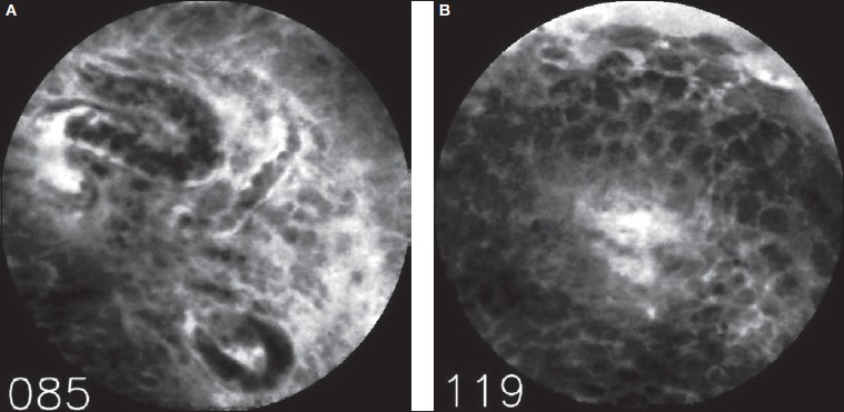
Example of a false-positive finding. Both examiners independently assessed this sequence as representing malignant changes. Note the relatively poor quality of this CLE image with three capillaries filled with erythrocytes (frame 085, Fig. A). The second image represents a later part of the sequence (frame 119, Fig. B) showing many small, similar cells without clear aberrant pleomorphism.