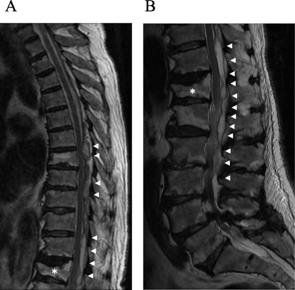
Fig. 1.

a, b Magnetic resonance imaging (T2) performed on admission. Fluid retention is observed in the epidural space behind the Th6-L3 spinal canal (arrow). Compression of the spinal cord near Th6/7, Th11/12, and L2/3 due to fluid retention is shown. Vertebral compression fractures can also be seen at Th12 (asterisk)
