
Fig. 2.
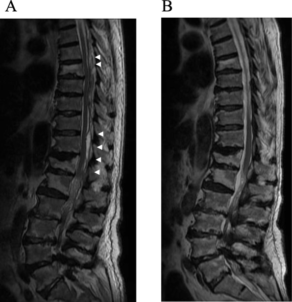
a Magnetic resonance imaging (MRI; T2) performed a day after fluoroscopy-guided percutaneous puncture. The volume of fluid in the epidural space has decreased and the spinal cord compression has been relieved (arrow). b MRI (T2) performed after 3 weeks. The epidural fluid pool has completely disappeared