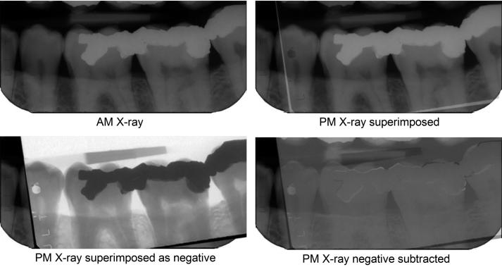
Figure 4.
Subtraction imaging. The postmortem (PM) image is superimposed on the antemortem (AM) image and turned into a negative. When its opacity is reduced, the AM image becomes progressively more visible until all the overlying negative and underlying positive colours cancel to resolve to grey wherever the images are similar. Changed features now become clear. The suggestion of a white margin around the amalgam fillings indicates a slight mismatch in the sensor or beam alignment or both between the two images, demonstrating that they are the same object. (Image courtesy Health Support Queensland Forensic and Scientific Services (HSQ FSS) and the Queensland State Coroner, with permission).

