
Figure 5.
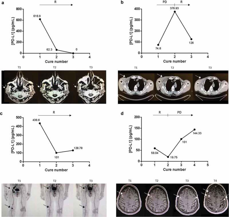
ExoPD-L1 can be used as follow-up markers in melanoma patients. Case study of the correlation between ExoPD-L1 levels in plasma samples from melanoma patients and response to PD-L1-based therapy. Concomitant imaging and ExoPD-L1 sampling in four patients experiencing (a) response as observed in the parotid gland and cervical lymph nodes on CT scan; (b) complete response in the subcutaneous tissue, popliteal and ilioinguinal lymph nodes on PET-CT; (c) disease response in the muscle and infraclavicular lymph nodes on CT scan; (d) initial response (at cure 1) then disease progression as detected in the brain on MRI. R: response, PD: progression of the disease. Tumour metastases in the scans are indicated by an arrow.