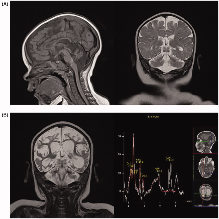
Figure 1.
(A) First MRI scan, age 5 months. Sagittal T1-weighted and coronal T2-weighted imaging showing diffuse white matter reduction, marked cerebral and cerebellar atrophy, corpus callosum thinning, and brainstem hypoplasia. (B) MRI scan, age 9 months. Coronal T2-weighted imaging showing marked progression of cerebral and cerebellar atrophy and ventriculomegaly (dominantly left occipital ventricle) due to diffuse white matter necrosis, while MRI spectroscopy showed markedly increased myoinositol peak, increased lactate peak, and reduced N-acetyl aspartate peak.

