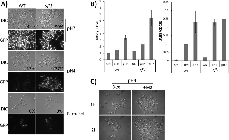
FIG 2.
(A) Morphology of WT and sfl1 strains expressing a copy of HWP1p-GFP after inoculation for 1 h in YPD medium set at pH 7 or pH 4 or supplemented with 100 μM farnesol. Percent filamentation is indicated at bottom right of DIC images. (B) qRT-PCR of BRG1 and UME6 transcripts after WT and sfl1 cells were grown at pH 4 and pH 7 for 1 h. qPCR values were normalized to CDC28 transcript levels for each sample. Presented data represent means ± SEM of results from 3 independent experiments. (C) Morphology of WT strain expressing a copy of MAL2p-BRG1 after inoculation for up to 2 h in YEP medium at pH 4 with either dextrose (+Dex) or maltose (+Mal) as the carbon source.

