FIG 1.
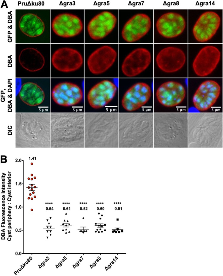
DBA staining intensity at the cyst periphery relative to that in the cyst interior is decreased in mutant strains that lack expression of PVM-associated GRA proteins. (A to B) In vitro cysts derived from different GRA deletion strains or the parental PruΔku80 strain were differentiated for 3 days. (A) Cysts were located using differential interference contrast (DIC) microscopy and imaged by confocal microscopy. The presence of bradyzoites inside cysts was verified by locating parasite nuclei with 4′,6-diamidino-2-phenylindole (DAPI) staining and verifying that each parasite nucleus was surrounded by expression of cytosolic green fluorescent protein (GFP) (GFP+ bradyzoites). Cysts were stained with Dolichos biflorus agglutinin (DBA). Panels show GFP and DBA; DBA; GFP, DBA, and DAPI; and DIC. Bar, 5 μm. (B) Cysts from each strain were analyzed to determine the ratio of DBA staining intensity at the cyst periphery relative to that in the cyst interior. Data were plotted as the average ratio mean of fluorescence intensity at the cyst periphery to the cyst interior ± standard error of the mean (SEM) for each strain. Δgra3 (n = 9), Δgra5 (n = 10), Δgra7 (n = 6), Δgra8 (n = 13), Δgra14 (n = 8), and PruΔku80 (n = 15). P values were calculated with a Student’s t test; ****, P < 0.0001.

