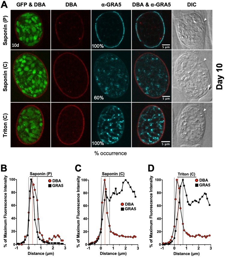
FIG 6.
Localization of GRA5 in 10-day-old mature cysts. (A) Infected HFFs on coverslips were treated under bradyzoite-inducing conditions for 10 days to differentiate mature in vitro cysts. Cysts were located using DIC microscopy and imaged by confocal microscopy. The presence of bradyzoites inside cysts was verified by locating parasite nuclei with DAPI staining (not shown) and verifying that each parasite nucleus was surrounded by expression of cytosolic GFP (GFP+ bradyzoites). Cysts fixed in 4% paraformaldehyde were permeabilized with either a short pulse (P) of saponin, with continuous (C) exposure to saponin, or with continuous (C) exposure to Triton. Cysts were stained with DBA and α-GRA5 antibody. Panels show GFP and DBA, DBA, α-GRA5, DBA and α-GRA5, and DIC (cyst wall indicated by white arrow). The percent occurrence is shown for GRA5 with saponin (P) (n = 14; 14/14 GRA5 at the cyst membrane/wall), saponin (C) (n = 10; 6/10 GRA5 not at the cyst membrane/wall), and Triton (n = 11; 11/11 GRA5 not at the cyst membrane/wall). Bar, 5 μm. (B to D) Fluorescence intensity profiles of representative cysts shown in panel A were generated to quantify the location of GRA protein(s) relative to the cyst wall, with DBA compared to α-GRA5 at day 10 for each method of permeabilization. The dotted black lines define the cyst wall region, and the dotted red line indicates the middle of the cyst wall, which corresponds to the peak DBA fluorescence intensity.

