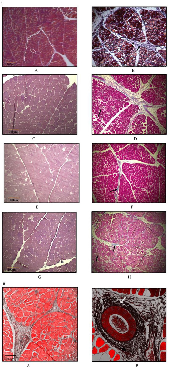
Figure 7.

Histomicrographs (Masson-Trichome staining) of cross-section of Pectoralis major. (i) Micrographs presented on left side (A,C,E,G) have less myodegeneration (non-myopathy birds) occurring in muscle fibers than micrographs (myopathy birds) presented on right side (B,D,F,H) for bird age of days- 21, 35, 42, and 57. Micrograph A: day 21 micrograph demonstrating tightly packed polygonal fibers with a minimal extracellular connective tissue space. White solid line outline represents the endomysial connective tissue space, and dashed line represents perimysial tissue space. Micrograph B: day 21 micrograph with wider perimysial region, and collagen deposition in perimysial region (indicated by arrow). (C,D) Day 35 micrographs; degenerating muscle fibers are elsewhere in micrograph D as indicated by black arrow and wider perimysial space filled with collagenous tissue (indicated by white arrow). (E,F) Day 42 micrographs; Perimysial and endomysial connective tissue spaces are wider in (F) as compared to (E) and the spaces are filled with collagenous tissue. Perivascular infiltration of immune cells was present around the blood vessels in (F) [magnified pic: (ii)]. (G,H) day 57 micrographs; Perimysial and endomysial connective tissue spaces filled with collagenous tissue [bluish coloration in (H)]. Greater variation in shape and size of fibers were observed in (H). Significant necrotic patches (white areas indicated by arrow) were also observed in degenerating fibers in (H). A tight parallel organization of perimysial collagen fibrils (indicated by arrow) are occurring as seen in (H) suggesting higher degree of collagen cross-linking. (ii) Histomicrographs of cross-section of P. major muscle at d 42 from a myopathy bird. Confocal images were obtained to improve contrast between vein, infiltrating cells and myofibers. (A) Thick perimysial collagenous tissue and fibrosed veins (indicated within white boxes) seen in connective tissue spaces leading to atrophy of peripheral fibers. (B) Magnified image of a fibrosed vein with perivascular infiltration of densely packed immune cells (indicated by white arrow).
