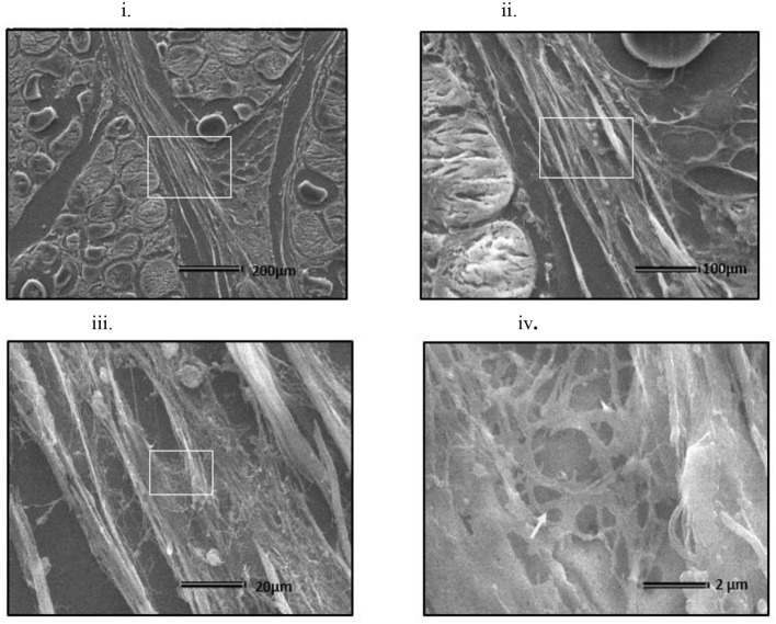
Figure 8.
Scanning electron micrograph of cross section of myopathy affected Pectoralis major muscle (i) Collagen fibrils making collagen fiber bundles (bundle width measured up to several μm in diameter) were evident and were tightly arranged across longitudinally in perimysial spaces. White arrows on image (i) indicates degenerating muscle fiber. White rectangular area on image (i) were sequentially magnified to get images (ii–iv) to obtain ultrastructure entangled collagen fibrils in image (iv) (indicated by white arrow).

