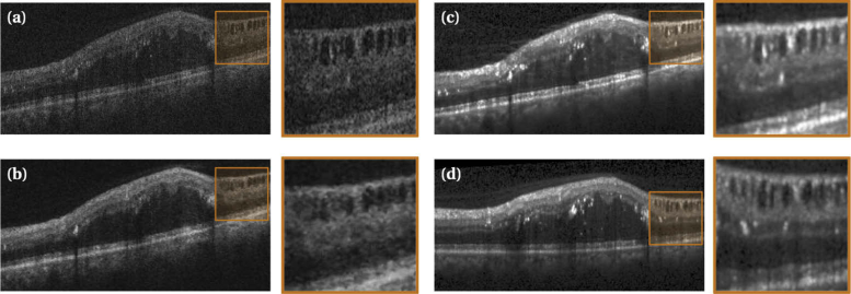
Fig. 3.
Qualitative results of the image translation algorithms. An original Cirrus OCT B-scan (a) was translated to the Spectralis domain using (b), and (c). The corresponding original Spectralis B-scan (d) acquired from the same patient at approximately the same time and retinal location is also shown for reference. The image generated in (c) has intensity values and an image noise level similar to those observed in the original Spectralis image (d).

