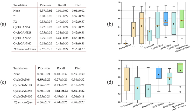
Fig. 7.
Quantitative results of sub-retinal fluid (SRF) segmentation, obtained on (a-b) Cirrus and (c-d) Spectralis scans. For different translation strategies Precision, Recall and Dice are shown together with the corresponding box-plots of Dice values. Native upper-bound models are indicated by * in (a-b). The CycleGAN model with highest mean Dice is highlighted in dark blue (b,d).

