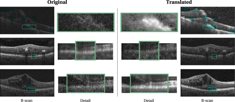
Fig. 11.
Left: Original B-scan and a zoomed-in region of interest ( original size). Right: Translated B-scan with the same zoomed-in region of interest. Regions that were identified as morphological differences by the experts are highlighted in cyan. First row: Spectralis-to-Cirrus translation. Second and third row: Cirrus-to-Spectralis translation.

