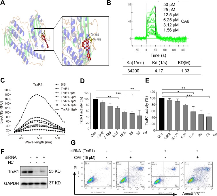
Figure 5.
CA6 binds to TrxR1 and suppresses its enzymatic activity. (A) Molecular docking between CA6 and TrxR1 was simulated with Tripos molecular modeling package Sybyl-x.v1.1.083. (B) Recombinant human (rh) TrxR1 was added to different concentration of CA6 and binding affinities were measured using SPR. (C) Binding between CA6 and rhTrxR1 was analyzed through bis-ANS displacement assay. (D) The inhibitory effect of CA6 on rhTrxR1 enzymatic activity was determined using DTNB assay [Mean ± SEM; n=3; **P<0.01, ***P<0.001]. (E) TrxR1 enzymatic activity in BGC-823 cell lysates was determined using end-point insulin reduction assay [Mean ± SEM; n=3; *P<0.05, **P<0.01, ***P<0.001]. (F) BGC-823 cells were transfected with siRNA against TrxR1 and levels of TrxR1 were determined via Western blotting. GAPDH was used as loading control. Representative immunoblot shown [n = 3]. (G) BGC-823 cells transfected with TrxR1 siRNA were exposed to CA6 (15 µM) for 24 h. Apoptotic cells were determined using Annexin V/PI staining. Representative dot plots from 3 independent experiments were shown.

