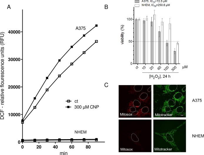
Fig 2. A higher basal (mitochondrial) ROS level in melanoma cells compared to melanocytes makes them more sensitive to exogenous H2O2.
A, ROS formation in presence and absence of CNP in melanoma cells and melanocytes was determined by relative fluorescence of DCF. B, Hydrogen peroxide (24 h) dose-dependently reduced the viability of both A375 and NHEM, but with a different IC50 value. C, A375 per se exhibited an increased level of mitochondrial superoxide (as indicated by the fluorescent dye MitoSOX (10 μM,10 min) compared to melanocytes. The fluorescent dye Mitotracker (100 nM) was used to visualize mitochondria, and to better distinguish between the cells, nuclei were encircled with a white dotted line.

