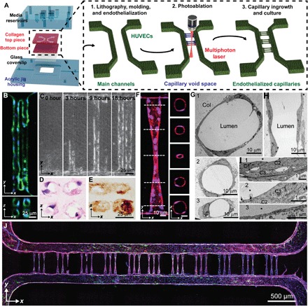
Fig. 1. Photoablation-guided capillary growth in lithography-based microvessel devices.

(A) Diagram of device assembly and capillary fabrication. Main channels were generated by soft lithography in acrylic jigs followed by capillary generation by photoablation and endothelial ingrowth. (B) Array (2 × 2) of 20-μm-diameter vessels demonstrates stable vessel lumens. Green: F-actin; blue: nuclei. (C) Endothelial ingrowth over 18 hours demonstrates complete vessel formation. (D and E) Cryosectioned capillaries were stained with (D) hematoxylin and eosin as well as (E) type IV collagen, demonstrating lumen formation by single endothelial cells and robust basement membrane deposition. (F) Constriction vessel design allows for the generation of sub–10-μm-diameter capillaries shown in both projected and cross-sectional views. Red: VE-cadherin; blue: nuclei. (G to I) Ultrastructural analysis of capillary vessel regions by transmission electron microscopy (TEM) shows vessels at varying diameters (from 40 to 10 μm) in cross-sectional (G and I) and longitudinal (H) views and varying wall thicknesses and junctions at cell-cell contact in zoomed views (I). Cross-sectional views of vessel regions near the connection. Col: collagen substrate; *: lumen; C: cells. (J) Stitched confocal image demonstrates complete vessel network consisting of 33 capillaries. Green: von Willebrand factor; blue: nuclei; red: VE-cadherin; purple: F-actin.
