
Fig. 5. Illustration of the self-cleaning of a contaminated superhydrophobic surface using confocal microscopy and friction force measurements.
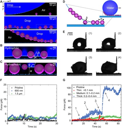
(A) A 10-μl water drop (dyed with ATTO 488; navy blue) is dragged over a nanoporous superhydrophobic surface contaminated with 10- to 50-μm hydrophilic particles (purple). The interface between the drop and the surface is monitored by LSCM. Particle contamination is completely taken along by the water drop (see Materials and Methods and fig. S9 for details of the image processing). (B and C) High-magnification LSCM images showing the contact angle θ of the hydrophilic and hydrophobic particles in contact with water. Smaller particles lost contact with the solid surface. (D) Sketch of the pickup process of particles. The deformed meniscus pulls on the particle. (E) Macroscopic observation of a 10-μl drop being dragged over a surface heavily contaminated with hydrophilic 10- to 50-μm particles. (F) Force required to clean a surface contaminated with hydrophilic 1.5-μm and 600-nm particles. The drop is moved at a velocity of v = 250 μm s−1. (G) Effect of the thickness of the contamination layer (<0.1 mm, 0.1 to 0.2 mm, and 0.2 to 0.4 mm) for hydrophilic 10- to 50-μm particles on the force required to clean the surface. For strongly contaminated surfaces (0.2- to 0.4-mm contamination layer), a continuous increase in the force during the self-cleaning can be observed (1 and 2). Upon complete coverage of the drop’s surface with particles (between 3 and 4), a sudden increase in force can be observed. Drop velocity, v = 250 μm s−1.