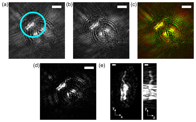
Fig. 6.
Example data acquired from a subject with heterogeneous scattering properties: a promastigote L. mexicana cell. (a) and (b) show the red and green maximum intensity projection images, respectively (scale bar = 10 μm in each). Panel (c) shows a color image with the red and green channels combined, so that the artifacts can be seen to lie in different positions in each channel. Panel (d) shows the maximum intensity projection of the registered ‘joint’ image (scale bar = 10 μm), and panel (e) shows two orthogonal projections of the cell, demonstrating the three-dimensional reconstruction of the flagellum, the hair-like projection at the top of the cell (scale bar = 2 μm).

