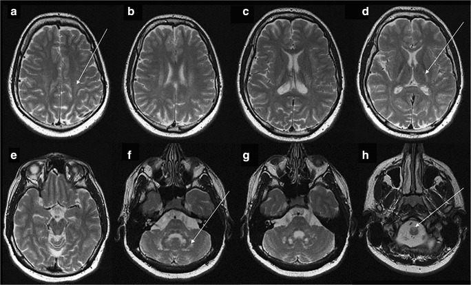
Fig. 1.
MRI examination at the age of 21 years. Axial T2-weighted images. See the bilateral hyperintense signal of the posterior part of the centrum semiovale (arrow on a), posterior limb of internal capsule (arrow on d), cerebellar white matter (arrow on f). The antero-medial part of medulla oblongata is also hyperintesive (arrow on h)

