Figure 2.
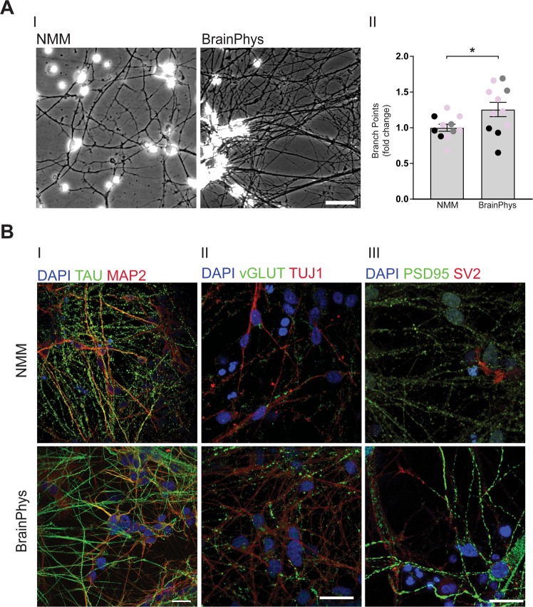
BrainPhys increases neuronal networks. NPCs were differentiated in BrainPhys or NMM for up to 35 days and investigated for changes in neuronal networks using bright-field imaging and immunocytochemistry. (A) BrainPhys increases neurite branching. (I) Representative bright-field images showing the neuronal networks in cells cultured in NMM (left panel) or BrainPhys (right panel). Scale bar = 20 µm. (II) Quantification of the number of neurite branch points shows an increase in cells cultured in BrainPhys already after ten days. Two-three images/experiment and condition from four separate experiments on neurons from three different iPSC lines (Ctrl1 marked with grey circles, ChiPSC22 marked with black circles and WTSIi015-A marked with pink circles) were analysed with Student’s t-test. *p ≤ 0.05. Bars represent mean +/− SEM presented as fold change relative to NMM. (B) BrainPhys increases markers of neuronal processes and synapses. Representative immunocytochemistry images of neuronal markers in NMM- and BrainPhys-cultured cells. (I) TAU (microtubule-associated protein tau, green): a marker of axonal microtubules and MAP2 (microtubule-associated protein 2, red): a marker for neuron-specific microtubuli of dendrites. (II) vGLUT1 (vesicular glutamate transporter 1, green): a synaptic marker for glutamatergic pre-synapses and TUJ1 (neuron-specific class III beta-tubulin, red): a marker for microtubule stability of axons. (III) PSD-95 (postsynaptic density protein 95, green): a marker of post-synaptic density and SV2 (synaptic vesicle protein 2, red): a marker of synaptic vesicles. Upper panel = NMM, lower panel = BrainPhys. Scale bar = 25 µm.

