Figure 5.
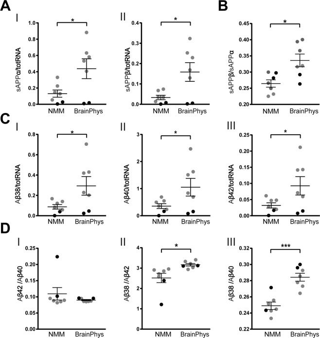
BrainPhys changes APP processing. NPCs differentiated in BrainPhys or NMM for up to 35 days and the concentrations of APP-cleavage products in the cell-conditioned media measured with immunochemiluminescence methods. Concentrations of secreted cleavage products of APP normalized to total RNA extracted from the corresponding cell lysate. (A) Concentrations of secreted sAPPα (I) and sAPPβ (II) measured in the cell-conditioned media and normalized to total RNA from the corresponding cell lysate, both significantly increase in BrainPhys-cultured neurons compared with NMM cultures. (B) The ratio of sAPPβ to sAPPα increases in BrainPhys-cultured cells compared with NMM cultures, indicating an increased β-site cleavage of APP. (C) Concentrations of secreted Aβ peptides were measured in the cell-conditioned media and normalized to total RNA from the corresponding cell lysate. Aβ38 (I), Aβ40 (II) and Aβ42 (III) all increase significantly with BrainPhys compared with NMM. (D) The ratios of Aβ42 to Aβ40 (I) does not differ between BrainPhys- and NMM cultures, whereas the ratio of Aβ38 to Aβ42 (II) and Aβ38 to Aβ40 (III) increase significantly. This indicates an increased secretion of Aβ38 over Aβ42 and Aβ40 in the BrainPhys-cultured cells. Mean values of seven separate experiments on neurons from two different iPSC lines (Ctrl1 marked with grey circles and ChiPSC22 marked with black circles) were analysed with Student’s t-test. *p ≤ 0.05, **p ≤ 0.01, ***p ≤ 0.001. Bars represent mean +/− SEM.

