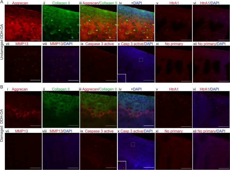
Figure 5.
Signs of remodeling but not of significant ECM degradation were detected in undamaged DDH-OA samples. (A) Panel showing an undamaged DDH-OA sample with MMS 2.5 stained for Aggrecan (i, red), Collagen II (ii, green), overlay (iii), overlay with DAPI (iv), HtrA1 (v, red), HtrA1 + DAPI (vi), MMP13 (vii, red), MMP13 + DAPI (viii), Caspase-3 active (ix, red), Caspase-3 active + DAPI (x) with inset for cells in higher magnification, No primary control (xi, red), and No primary control + DAPI (xii). Only Aggrecan and Collagen II were co-stained on the same sample. The images for the other antibodies’ staining are in equivalent areas. (B) Same array of panels for a damaged DDH-OA sample with MMS 4.5. Scale bars are 200 µm. Images were taken with 20x objective.

