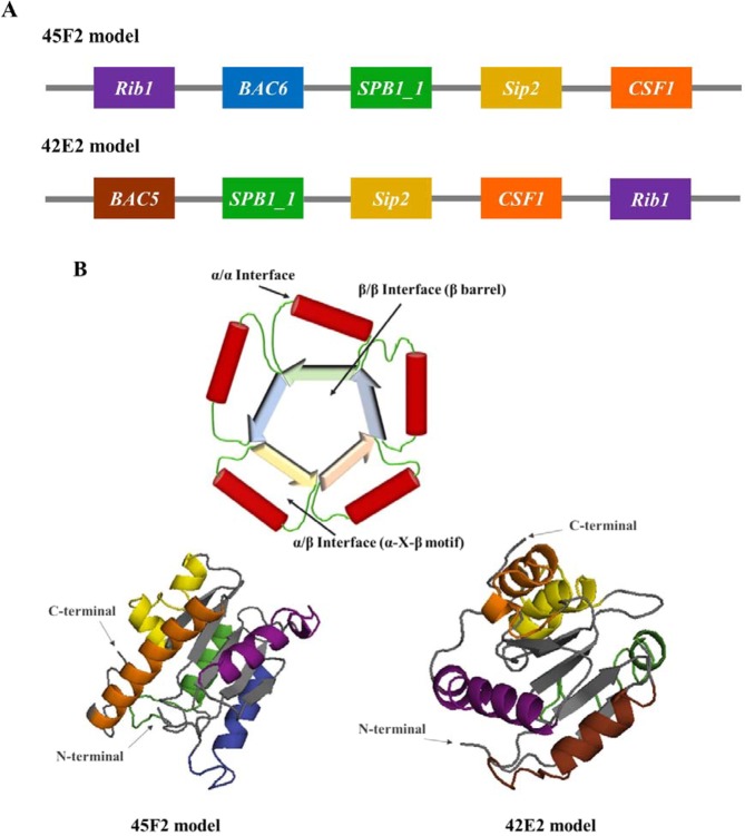
Figure 3.
Schematic diagram and predicted 3D model of designed chimeric multiepitope vaccines. (A) The schematic diagram depicting the chimeric multiepitope vaccine of the 45F2 and 42E2 constructs containing 5 different B cell epitopes in α-helical structures (Rip1, BAC6, BAC5, SPB1_1, Sip2, and CSF1 illustrated in violet, blue, brown, green, yellow and orange, respectively) linked with 6 fragments of a β-pleated sheet of the flavodoxin backbone (gray color). (B) The predicted 3D protein structures of the 45F2 and 42E2 models, which were the two best vaccine candidates, are shown as α/β proteins with a flavodoxin fold. Their colors are represented as colors in the schematic diagram, and their N terminus and C terminus are indicated by arrows. These vaccines were designed based on the desirable construction of the TIM-barrel structure, as shown in Fig. 4B.

