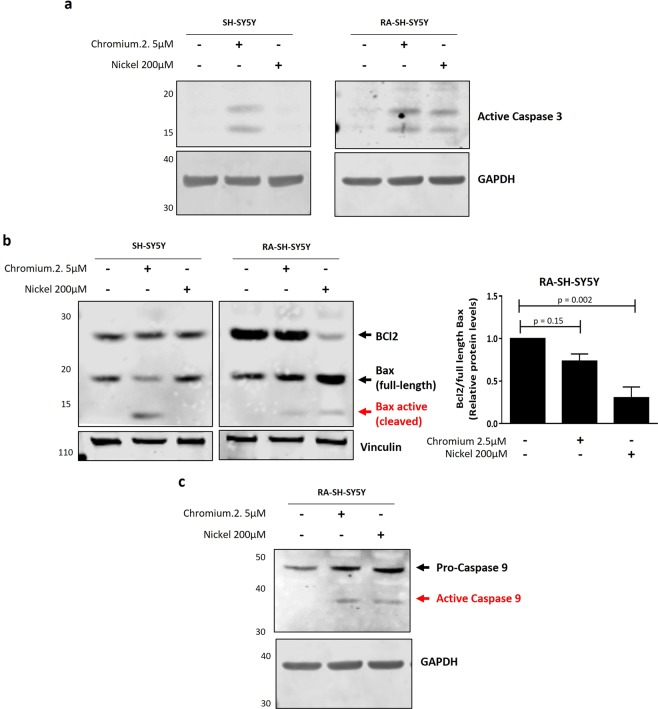
Figure 5.
Cr and Ni exposure induces apoptotic cell death in SH-SY5Y cells. Non-differentiated and RA-differentiated SH-SY5Y cells were treated with Cr (2.5 µM) and Ni (200 µM) for 24 hours. Whole cell lysates were collected in order to analyze by western blot the levels of cleaved caspase-3 protein (a), the ratio of anti-apoptotic protein Bcl2 to pro-apoptotic protein Bax (b) and the cleavage and activation of caspase-9 (c). Vinculin and GAPDH were used as loading controls. (a) Representative immunoblot showing the presence of the 17/19KD fragment of caspase-3 after Cr and Ni exposure. (b) Representative immunoblots showing the levels of Bcl2 and Bax proteins after heavy metal exposure. The activation of the intrinsic/mitochondrial apoptosis pathway was assessed by the decrease of the ratio of Bcl2 to full length Bax and the presence of a cleavage form of Bax protein. The plot represent the average ± SEM of 3 independent experiments. Statistical significance was determined by one-way analysis of variance (anova) followed by Bonferroni’s test for multiple comparisons using GraphPad Prism 6. p-values comparing untreated and heavy metal treated cells are included. Statistical significance was considered when p-value ≤ 0.05. (c) Immunoblot showing the presence of the fragment (43KD) of active caspase-9 after Cr and Ni exposure to confirm the activation of the intrinsic/mitochondrial pathway. All experiments were performed in triplicates.

