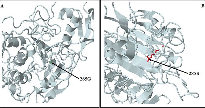
Figure 2.

Predicted structure of the N-terminal region structure of SMCHD1 showing the amino acid change resulting from c.853G>C. The predicted models are based on the template c5ix1A (PDB header: transcription; PDB molecule: MORC family CW-type zinc finger protein 3; PDBTitle: crystal structure of mouse Morc3 ATPase-CW cassette in complex with AMPPNP and H3K4me3 peptide). (A) SMCHD1 structure showing the wild-type residue (G). (B) SMCHD1 structure showing the variant residue (R).
