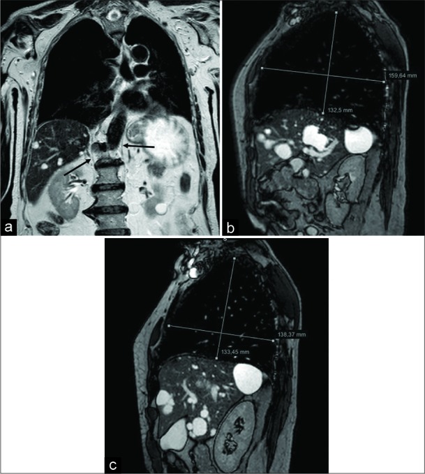
Figure 4:

A 76-year-old male patient affected by Pompe disease. Coronal turbo spin echo T2-w acquisition (a) shows and advanced state of hypotrophy of both the diaphragmatic crura (arrows). Sagittal balanced-fast field echo scans, performed at end-inspiration (b) and end-expiration (c), demonstrate a slight lung volume change, mainly appreciable on the anteroposterior plane, due to both the decreased function of the diaphragm and the partial intercostal muscles compensation.
