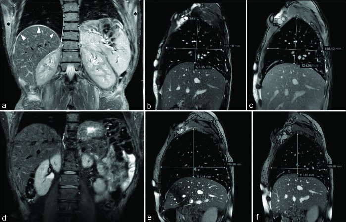
Figure 5:
A 45-year-old male complaining lower limb pain and weakness as well as shortening of the breath. Magnetic resonance imaging demonstrated polymyositis. Coronal fast T2-w STIR scan (a) shows hyperintensity of the right hemidiaphragmatic dome (arrowheads) and upper position of the left diaphragm due to inflammation. Note the presence of a bilateral hyperintensity of the chest wall muscles (asterisks). Sagittal balanced-fast field echo scans (acquisition time: 0.6 s) passing through the middle of the right diaphragm, acquired in deep inspiration (b) and deep expiration (c), demonstrate complete absence of diaphragmatic movement. Coronal T2-w STIR performed 3 months after corticosteroid treatment (d) shows the disappearance of the diaphragmatic and chest muscle edema. On sagittal balanced-fast field echo scans obtained during inspiration (e) and expiration (f), a partial recovery of the movement can be detected. The patient had a significative, although partial, improvement of the respiratory symptoms.

