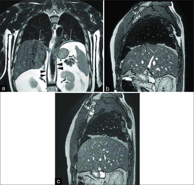
Figure 6:

A 50-year-old male patient with respiratory failure of unknown origin. Computed tomography-scan showed bilateral basal bands of parenchymal atelectasis. The functional examination was suspicious for conduction abnormalities of both the phrenic nerves. Coronal turbo spin echo T2-w image (a) shows normal diaphragmatic crura (arrowheads), upper position of both the hemidiaphragms and disventilatory lung parenchyma atelectasis (arrows). Inspiratory (b) and expiratory (c) sagittal balanced- fast field echo scans demonstrate the absence of diaphragmatic movements, a slight increase of the anteroposterior diameter of the lung can be seen during inspiration due to the contraction of the intercostal muscles. The final diagnosis was bilateral phrenic nerve paralysis due to polyneuritis.
