Fig. 5.
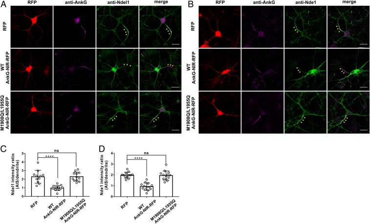
Overexpression of an AnkG-NIR peptide abolished AIS enrichment of Ndel1 and Nde1. (A and B) Neurons transfected with RFP control, WT AnkG-NIR-RFP, or M1908Q/L1955Q AnkG-NIR-RFP and stained for AnkG (magenta) or endogenous Ndel1 (A) or Nde1 (B). Yellow arrowheads indicate the AIS. (Scale bars: 20 μm.) (C) Quantification of the Ndel1 fluorescence intensity ratio of the AIS to dendrites in neurons transfected with RFP (n = 11), WT AnkG-NIR-RFP (n = 13), or M1908Q/L1955Q AnkG-NIR-RFP (n = 12). Error bars indicate SE. ****P < 0.0001. (D) Quantification of the Nde1 fluorescence intensity ratio of AIS to dendrites in neurons transfected with RFP (n = 12), WT AnkG-NIR-RFP (n = 13), or M1908Q/L1955Q AnkG-NIR-RFP (n = 13). Error bars indicate SE. ****P < 0.0001; ns, not significant.

