Fig. 6.
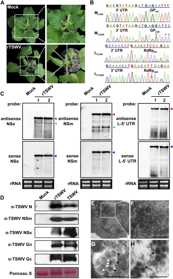
Rescue of infectious TSWV from full-length cDNA clones in N. benthamiana. (A) Systemic infection of N. benthamiana plants with rescued TSWV (rTSWV) resulting from agroinfiltration of S(+), M(−)opt, and L(+)opt and 3 VSRs (P19, HcPro, and γb). The plant agroinfiltrated with pCB301 empty vector was used as a mock control. Images were taken at 19 dpi. Boxed areas (Left) of the plants that show stunting, mosaic, and leaf curling are shown enlarged (Right). (B) Sequence confirmation of codon-optimized sequences of GP gene [from M(−)opt RNA segment] and RdRp gene [from the L(+)opt RNA segment] on RT-PCR fragments obtained from systemic leaves of N. benthamiana infected with rTSWV. The optimized sequence of GP from rTSWV is underlined with a blue dashed line; wild-type GP sequence is underlined in blue. The 3′-untranslated region (UTR) sequence of the M genomic RNA is marked with a yellow line. The optimized sequence of RdRp from rTSWV is underlined with a red dashed line, and wild-type RdRp sequence is underlined in red. The 3′-UTR sequence of the L genomic RNA is marked with a purple line. The stars indicate codon-optimization sites of GP and RdRp gene sequences. (C) Northern blot detection of viral RNA from the S, M, and L RNA segment, respectively, in systemically leaves of N. benthamiana infected with rTSWV. Genomic RNAs (red arrow), antigenomic RNAs (blue arrow), and subgenomic RNAs (green arrow) were detected with DIG-labeled sense and antisense NSs-, NSm-, and L-5′ UTR probes, respectively. Lanes 1 and 2 refer to 2 independent replicates. Ethidium bromide staining was used as RNA loading control. (D) Western immunoblot detection of the viral proteins from leaves systemically infected with rTSWV using specific antibodies against N, NSm, NSs, Gc, and Gn, respectively. Leaves infected with wild-type TSWV were used as a positive control. Ponceau S staining was used as protein loading control. (E and F) Electron micrographs of thin sections of N. benthamiana plants infected with rTSWV (E). Boxed regions (E) show virions and are also shown enlarged (F). (G) Immunogold labeling of spherical, enveloped virus particles with anti-serum against Gn followed by a goat anti-rabbit Immunoglobulin G (IgG) conjugated with gold particles. (H) Immunogold labeling of virus particles using only the goat anti-rabbit IgG conjugated with gold particles. White arrowheads indicate gold particles; black arrowheads indicate spherical, enveloped virus particles. (Scale bars: 0.2 μm.)

