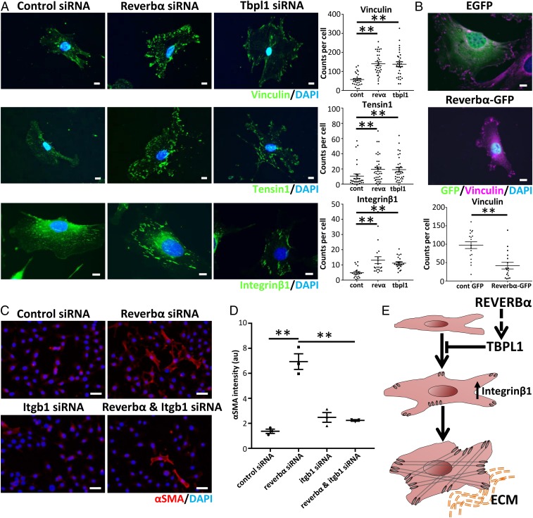
Fig. 4.
REVERBα and TBPL1 affect myofibroblast differentiation through changes in integrinβ1 expression. (A) Representative immunofluorescent images and quantification per cell of vinculin, tensin1, and integrinβ1 following siRNA knockdown of Reverbα or Tbpl1 compared to control (nontargeting) siRNA in mLF-hT cells (n = 3 separate transfections). **P < 0.01 (1-way ANOVA post hoc Dunnett test; mean ± SEM). Dots represent individual cells from 3 transfections. cont, control siRNA; revα, Reverbα siRNA. (Scale bars, 10 µm.) (B) Representative immunofluorescence image after mLF-hT cells have been transfected with REVERBα-GFP plasmid or an empty-GFP plasmid. Cells were stained for GFP, vinculin, and nuclei (4′,6-diamidino-2-phenylindole [DAPI]) (n = 3 separate transfections). **P < 0.01 (Student t test; mean ± SEM). Dots represent individual cells from 3 transfections with the focal-adhesion number being quantified per cell. (Scale bars, 10 µm.) (C) Representative immunofluorescence images. (Scale bars, 50 µm.) (D) Quantification of the myofibroblast marker αSMA following dual siRNA knockdown (control or Reverbα in the presence or absence of Itgb1) in mLF-hT cells (n = 3 separate transfections). **P ≤ 0.01 (1-way ANOVA post hoc Dunnett test; mean ± SEM). (E) Schematic demonstrating how both REVERBα and TBPL1 regulate Integrinβ1, which in turn affects myofibroblast differentiation. ECM, extracellular matrix.

