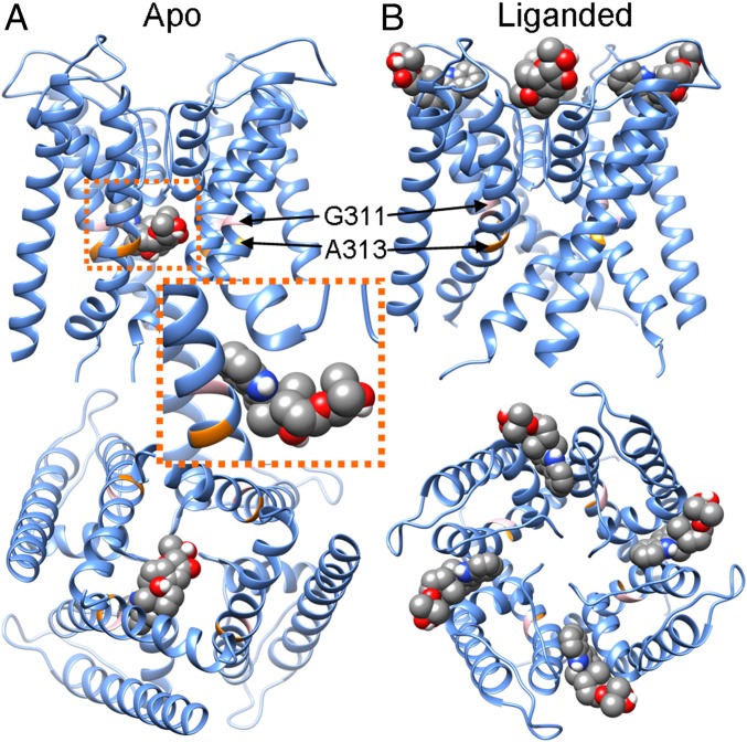
Fig. 1.
The best docking poses of PAX in the pore region of the closed and open mSl1o homologous structures. (A) The best docking pose of PAX in the PGD of the closed mSlo1 structure. For clarity, only 1 of 4 identical binding poses is shown, as these poses partially overlap. PAX is rendered in spheres, with carbon, oxygen, nitrogen, and hydrogen shown in gray, red, blue, and white, respectively. (Upper) Side view. (Lower) View from the intracellular entrance of the BK channel. G311 and A313 are shown in pink and orange, respectively. (Inset) Close-up view of PAX in the closed BK pore. (B) The best docking pose of PAX in the open mSlo1 pore. (Upper) Side view. (Lower) View from the extracellular entrance of the BK channel.

